This is a large group of conditions, and several are commonly seen in veterinary species. Congenital conditions tend to be evident at, or soon after, birth, but are not necessarily inherited. Inherited conditions indicate an underlying genetic etiology, but do not necessarily mean that the parents are also affected.
Primary central nervous system conditions
Muscles of the developing embryo require normal innervation in order to develop properly. Lack of normal innervation can lead to atrophy or replacement with fibrous connective tissue. Recall that motor neurons release trophic factors at the motor end plate that are critical to the health of skeletal muscle . Conditions that primarily affect the motor neurons, therefore, can have significant effects on skeletal muscle.
Arthrogryposis
Arthrogryposis refers to the abnormal angulature of the limbs (arthro: joint; gryposis: abnormal curvature). As you might expect, this condition is evident immediately at birth, and can affect one or more limbs in a multitude of different ways: they may be rotated, curved backwards or forwards, or abducted. The curve of the vertebral column may be affected as well: scoliosis (lateral deviation), kyphosis (dorsal deviation), or torticollis (twisted neck) are not uncommon. Muscle mass is reduced.
The causes of arthrogryposis are varied but are often unclear. There is a distinct association with dysraphism – the arrest or delayed closure of the neural tube (e.g. spina bifida) – and arthrogryposis. Other causes include toxins (e.g. wild lupine) and a variety of viruses, including the Orthobunyaviruses (Schmallenberg, Cache Valley, and Akabane viruses), bluetongue virus, and border disease virus.
Muscular defects
Splayleg
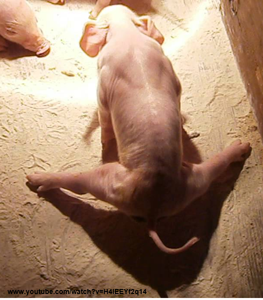
Splayleg is an odd condition that occurrs only in neonatal piglets (Figure 3.1). Piglets are born unable to adduct their limbs (particularly the hindlimbs) and are often found in a characteristic “splayed” position. The cause is unknown, but is thought to be associated with immature skeletal muscle present at the time of birth. Importantly, the condition is transient: all animals make a full recovery within 1, or at most 2, weeks. While affected, however, the animals are at risk of accidental injury and may become malnourished due to difficulty in nursing.
Muscular hyperplasia
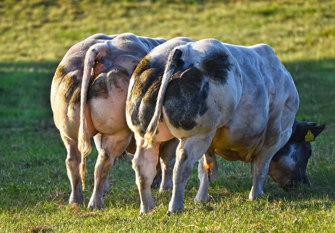
Recall that normal muscle cannot undergo hyperplasia: increase in muscle size is usually a function of hypertrophy. However, myofiber hyperplasia is the hallmark of a defective protein known as myostatin. Myostatin is a protein produced and released by myofibers and normally inhibits muscle growth. Genetic defects in the myostatin gene, resulting in dysfunctional myostatin, result in hyperplasia of skeletal muscle, known as “double muscling”. As this is hyperplasia, muscles have increased numbers of structurally normal myofibers. This trait has been selected for in several beef breeds, including Belgian Blue (Figure 3.2) and White. The condition is also seen in Whippet dogs (“bully” Whippets). Muscular hyperplasia is most pronounced in the thighs, rumps, loins, and shoulders.
Steatosis
Steatosis of muscles refers simply to the replacement of myofibers by adipocytes, usually following damage or denervation. It is generally considered an incidental finding at necropsy, but sometimes raises concern at meat inspection.
Congenital diaphragmatic clefts
Abnormal closure of the developmentally complex diaphragm can result in congenital diaphragmatic hernias. They have been reported most frequently in the dog and rabbit.
Muscular dystrophies
Muscular dystrophies (as defined in the human literature) are inherited conditions characterized by progressive myopathy with necrosis and regeneration of myofibers. Be aware that the term dystrophy is commonly misused in veterinary medicine, and only a few true dystrophies have been described, notably in the dog, cat, and sheep. These conditions typically manifest in young animals and progressively worsen as the animal ages. The canine and feline X-linked dystrophies are analagous to Duchenne and Becker muscular dystrophies of humans. Both are related to a mutation in the gene encoding dystrophin, a cytoskeletal protein. The pathogenesis of muscle damage in both conditions is poorly understood.
X-linked dystrophies of dogs and cats
The X-linked dystrophies of dogs and cats share several similarities and a few key diferences, summarized below in Table 3.1. Both conditions affect the dystrophin protein, whose exact function is still somewhat uncertain. Because affected gene is found on the X chromosome, males are overrepresented. Note that the level of detail presented here is beyond what you would be expected to know on an exam.
Ovine muscular dystrophy
Unlike the X-linked dystrophies of dogs and cats, ovine muscular dystrophy is an autosomal recessive condition that affects males and females equally. The condition is still common in Merino sheep in Australia, with 1-2% of animals showing signs of the disease. Clinical signs include a lack of normal growth, abnormal gait, and/or stiffness. Most animals will show clinical signs by 1 year of age, and be severely affected by 2-3 years old. If left unattended at pasture, severely affected animals are so weak that they die of starvation.
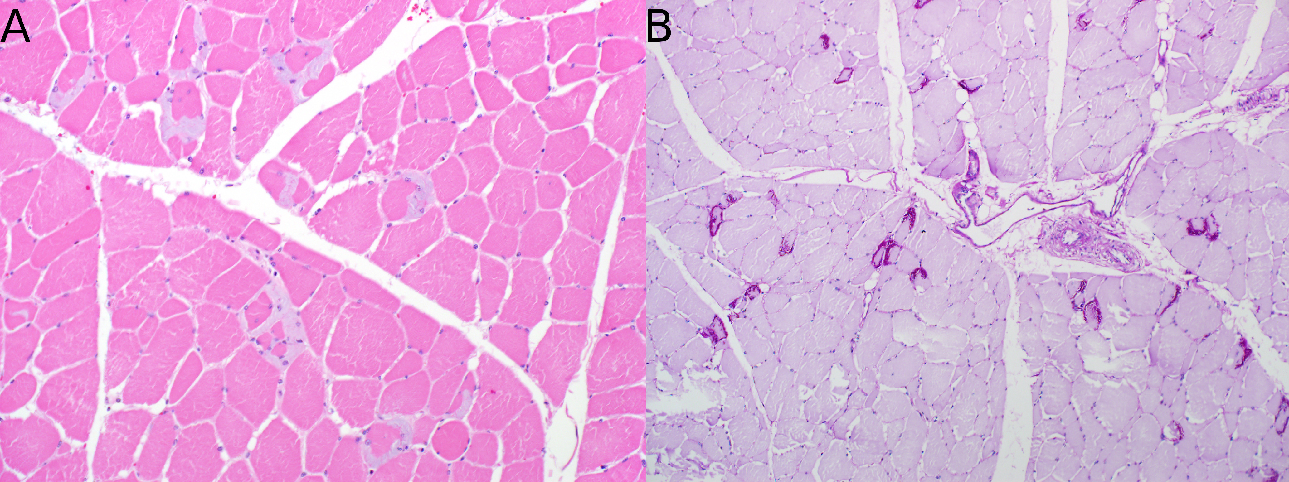
Gross changes include emaciation and replacement of muscles with adipose tissue. Histologically there are characteristic amphophilic sarcoplasmic masses and large, vesicular nuclei.
Malignant hyperthermia
Though clasically thought of as a disease of pigs, malignant hyperthermia is better thought of as a syndrome that predominantly occurs in swine, but which also affects dogs and horses. The underlying issue is a defect in the RYR1 gene, which encodes the ryanodine receptor, a calcium channel present within the sarcoplasmic reticulum. Defective ryanodine receptors are responsible for the release of Ca2+ during contraction. Defective ryanodine receptors stay open for longer, leading excess Ca2+ release, prolonged contraction, hypercontraction, and hyperthermia.
Porcine stress syndrome
Malignant hyperthermia is best characterized in pigs, and is also known as porcine stress syndrome (PSS). It is the cause of pale, soft, and exudative pork that is sometimes encountered at slaughter. A single nucleotide polymorphism in the RYR1 gene of pigs is the cause. It is estimated that between 2-30 % of pure-bred pigs are susceptible to PSS.
Pigs may be unaffected or subclinical. Stressful events (such as fighting or transport), or halothane anesthesia, however, can trigger severe episodes in which pigs display intense limb and torso rigidity, hyperthermia, tachycardia, dyspnea, metabolic acidosis, and rapid death. Gross lesions are related to the increased body temperature: classically pale, soft, exudative muscle (meat). Lesions attritutable to heart failure, including pulmonary edema, hydrothorax, and hepatic congestion, may occur if the animal survives the acute episode. Histologically, multifocal, monophasic necrosis is present, and edema separates myofibers in animals that have suffered from hyperthermia.
Congenital myasthenia gravis
Myasthenia gravis is a neuromuscular disorder characterized by decreased availability of acetylcholine (Ach) receptors (Ach-R) on the myofiber membrane. Lack of the receptor leads to decreased Ach binding, fewer depolarizations, and weaker muscle contractions. In congenital myasthenia gravis, decreased Ach-R availability is due to an inherent defect in the receptor. In [acquired myasthenia gravis][Acquired myasthenia gravis], circulating autoantibodies bind to and block the binding of Ach to their receptor. The acquired form is much more common than it’s congenital counterpart.
In dogs, congenital myasthenia gravis is autosomal recessive. Puppies develop exercise-induced collapse by 5-16 weeks of age, depending on breed. Regurgitation secondary to megaesophagus is a common clinical finding. Antibodies against Ach-R cannot be demonstrated.
The disorder is even less common in cats, which may seem normal at birth, but by approximately 4-5 months of age show signs of episodic weakness. Megaesophagus is not a feature. The condition remains poorly characterized.
Myotonic and spastic syndromes
Myotonia is the temporary inability of muscles to relax. It can be acquired or inherited; in veterinary species, the inherited form is most common. There are a variety of myotonias in dogs, cats, and goats that will not be covered here; more information can be found in the reference textbooks.
Hyperkalemic periodic paralysis (HYPP)
Only horses descended from the Quarter Horse stallion “Impressive” are at risk for HYPP. Affected animals have very well defined musculature, a desireable trait, which has led to the propogation of the condition in the quarter horse breed.
The pathogenesis of the condition is poorly understood. The underlying cause is a defect in Na+ channels causing excess influx of Na+ and efflux of K+ from myofibers. This alters the membrane potential of myofibers and leads to increased and prolonged action potentials, and excess contractile activity.
The condition presents differently depending on whether the animal is a heterozygote or homozygote. Homozygote foals characteristically suffer from laryngospasm, which can generate a distinctive inspiratory noise. They may develop dysphagia and become emaciated during their first 2 years. The larynx may collapse with exercise. Homozygotes typically do not survive beyond 2 years of age.
Heterozygotes are less severely affected, and often appear normal but suffer from transient attacks of muscle fasiculations and spams (myotonia), inspiratory stridor, protrusion of the third eyelid, and sometimes followed by acute paralytic collapse. Sudden death may occur in the most severe cases.
There are no gross or histologic lesions; diagnosis is based on ancestry, clinical history, signs, and DNA testing. Despite the name, hyperkalemia is not a consistent finding.